


Details
A mammogram, helps in the early detection and diagnosis of breast diseases in women. The mammography exam is specialized medical imaging that uses a low-dose x-ray system to see inside the breasts.The American Cancer Society recommends that every woman have her baseline (initial) mammogram at age 35, and begin having annual screening mammograms at age 40. WPG has now partnered with MOGA to make this screening tool more accessible to you
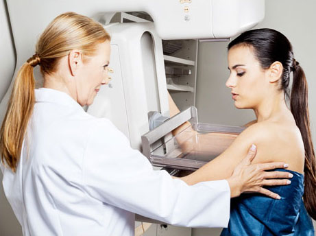
The technician will ask you to undress from the waist up, and put on a hospital gown which opens in the front. She will then position you next to the machine, helping you to rest each breast on a metal plate and arrange as much breast tissue as possible between the bottom plate and the top compression plate. The compression will last about ten seconds. During this time, the machine will make both vertical and lateral images. Sometimes, images will need to be re-done for better accuracy. This is simply an effort to get the most accurate results possible, and does not necessarily mean there is a problem.
Breasts are a tender part of your body, and the squeezing by the machine is – although necessary—somewhat uncomfortable. The good news is that there are some things you can do to lessen the discomfort:
Your results will be interpreted by a radiologist. The radiologist will often write a report and send it to your physician. You should also receive a letter by mail giving you the general results. If you do not hear from us within thirty days, please call and let us know. If your results are abnormal, remain calm. Only about 10% of women with abnormal screenings will truly have breast cancer. A digital mammogram or breast biopsy may be necessary for additional study.
3D mammograms, also known as tomosynthesis or “tomo”, use the same x-ray technology as “regular” 2D mammograms. Although the 3D mammogram takes a few seconds longer than the 2D, the experience for the patient feels the same. In both 3D and 2D mammograms, the breast is compressed between two plates. 2D mammograms take images only from the front and side views, which may create images with overlapping breast tissue. However during a 3D Mammogram, an x-ray arm sweeps over and takes additional images of the breast which minimizes overlapping breast tissue and gives radiologists the ability to more clearly view the fine details inside the breast, layer by layer.